


Invitro Diagnostics - medical services in Moldova


We are in numbers
0
laboratory tests
0
doctors
0
years in the industry
0
points on the map
0
results in 1 day
0
patients


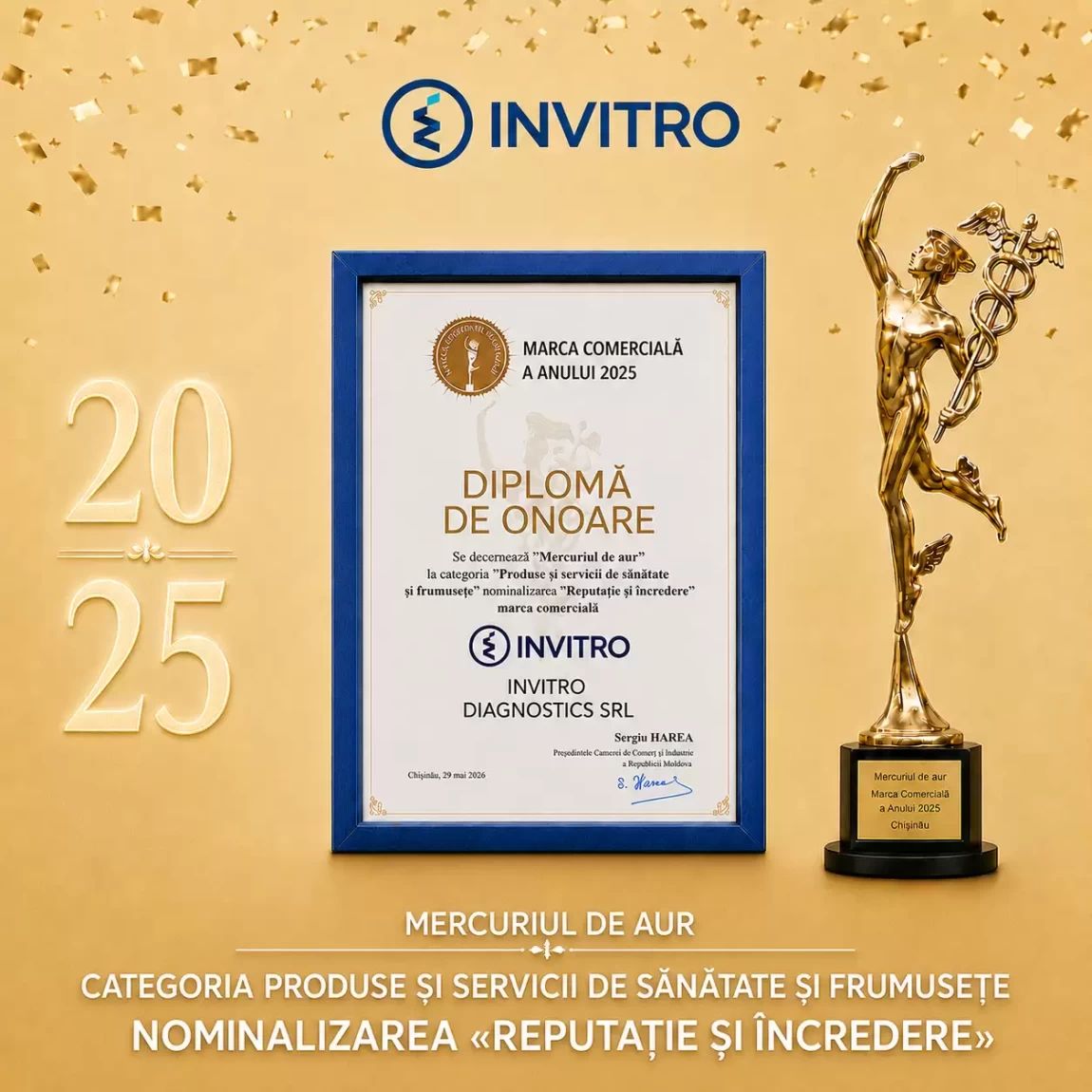
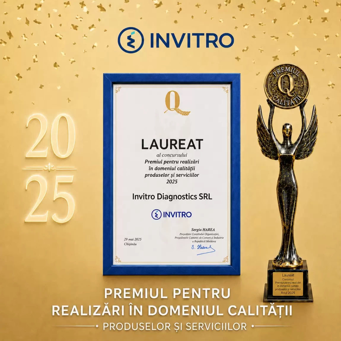
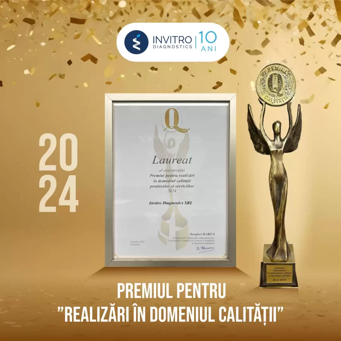
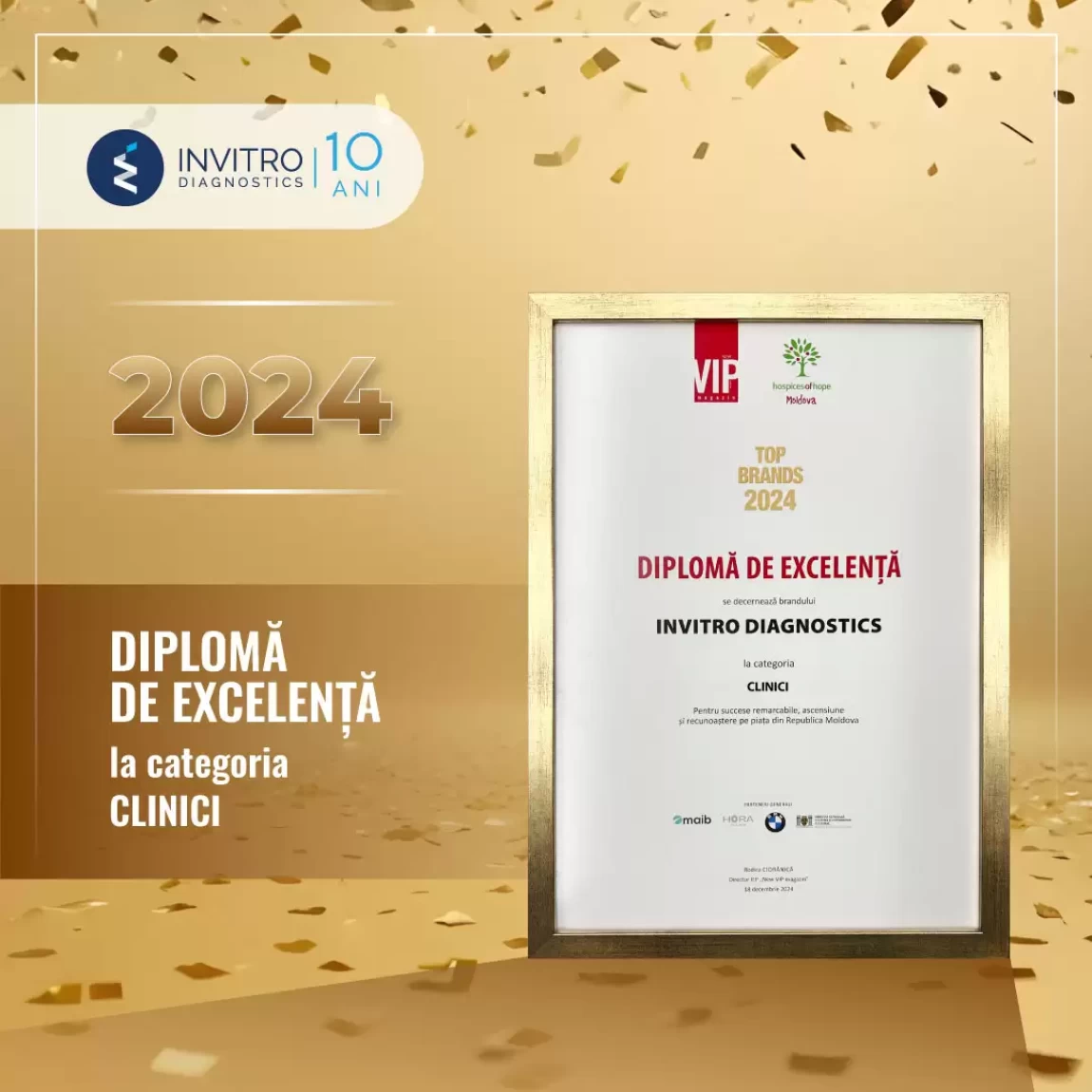
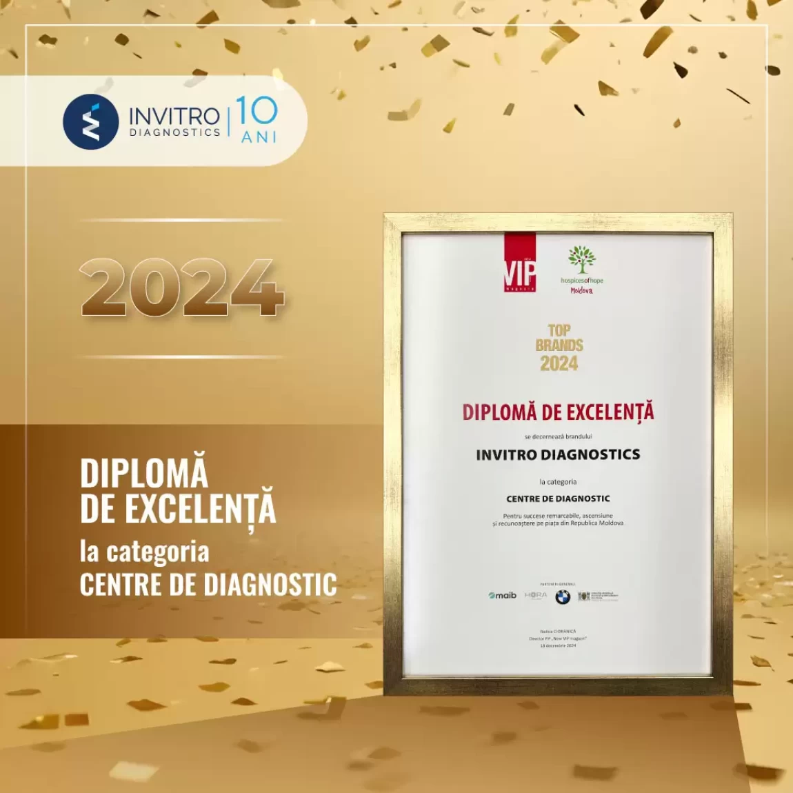
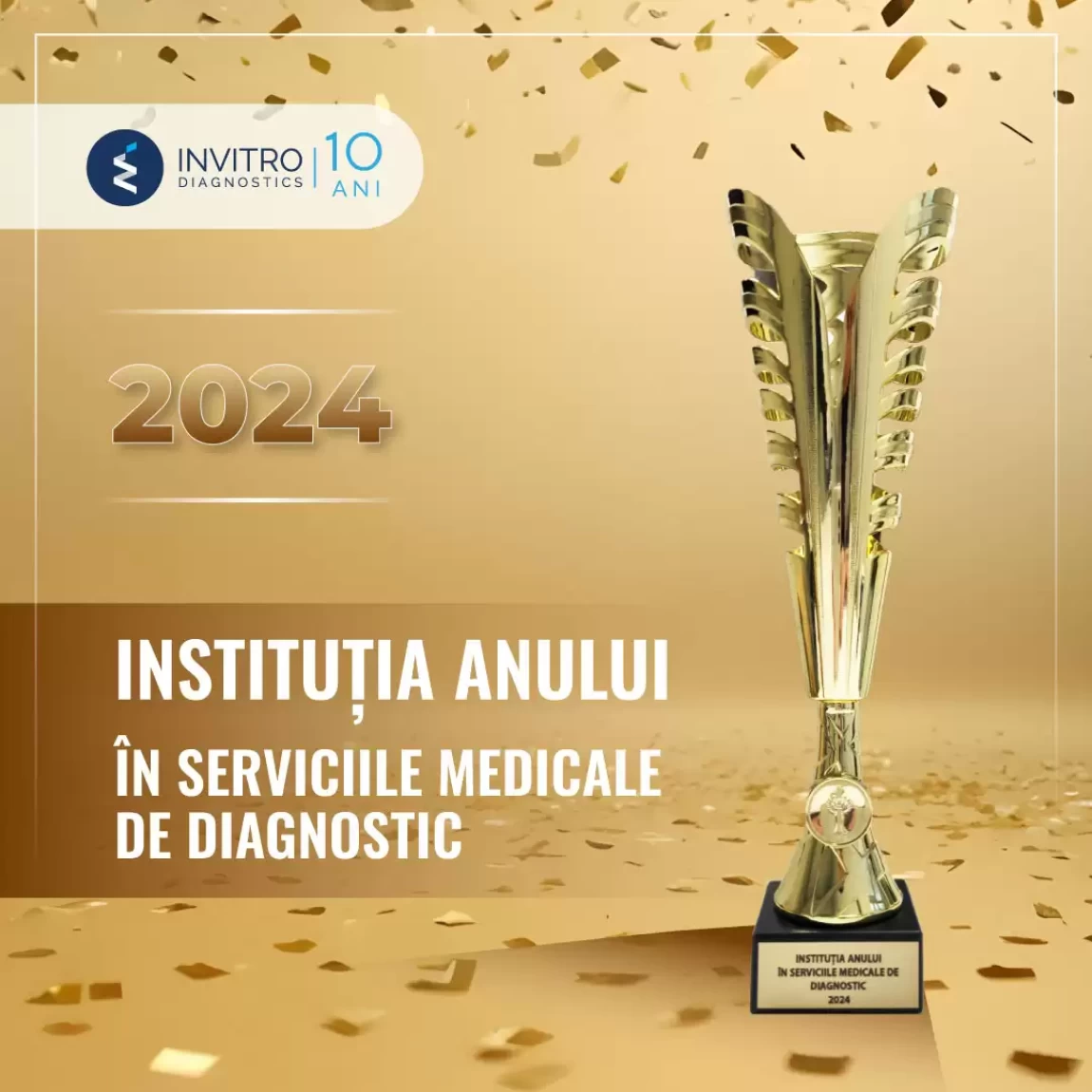
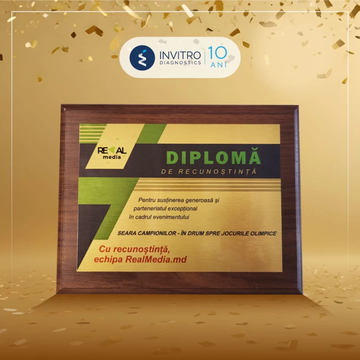
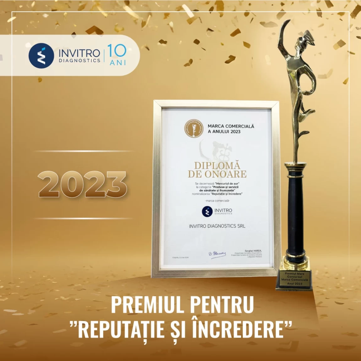
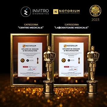
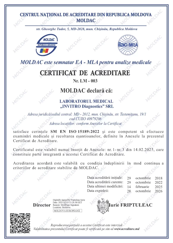
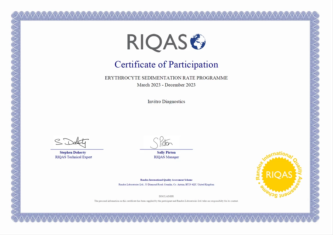
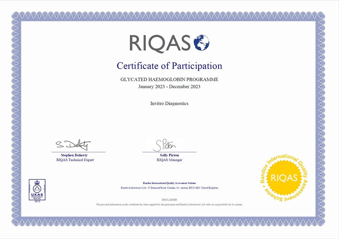
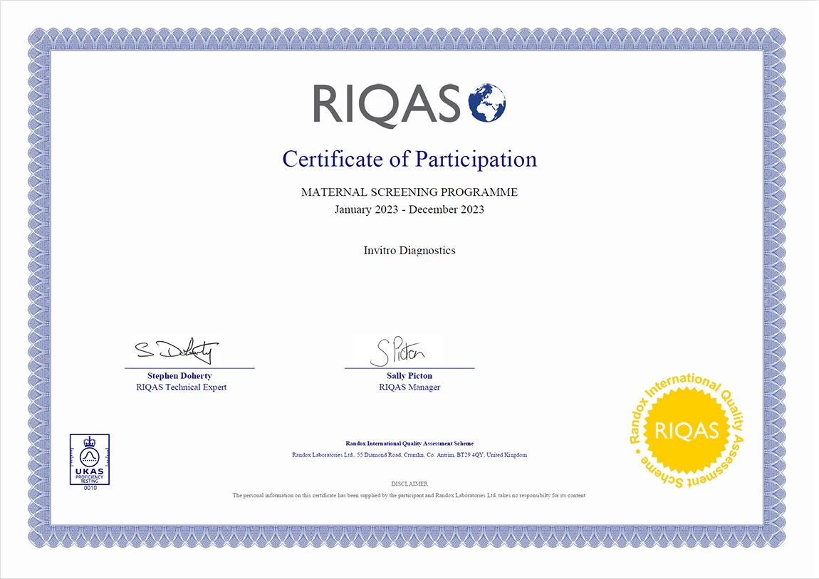
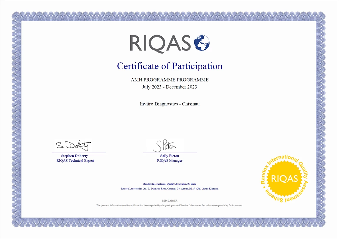
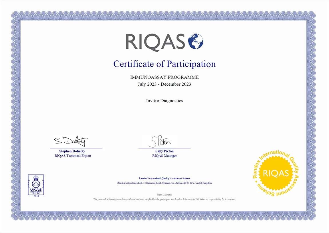
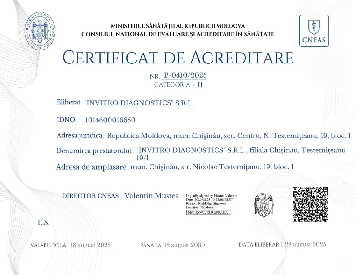
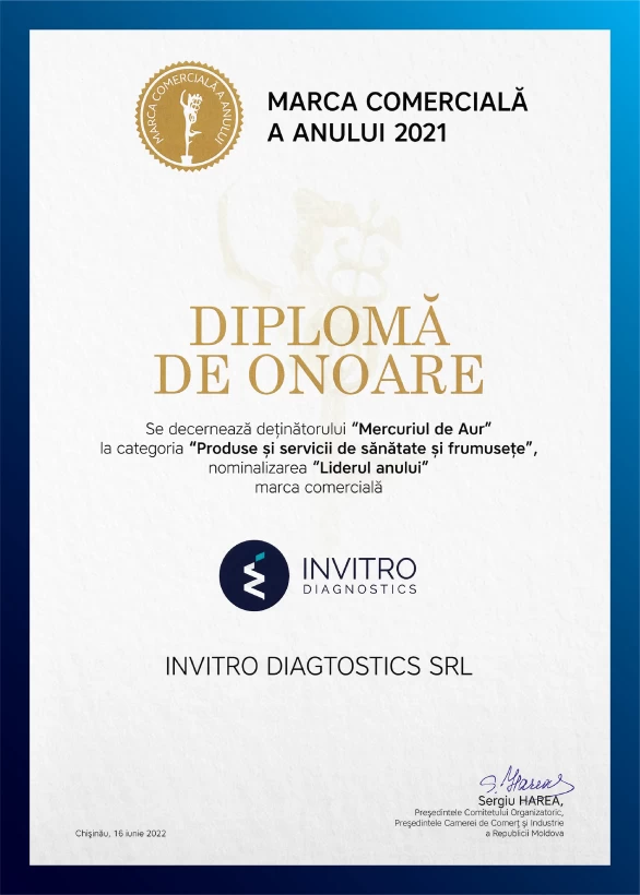
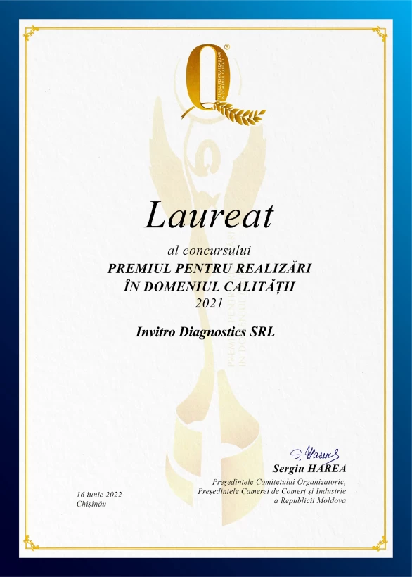
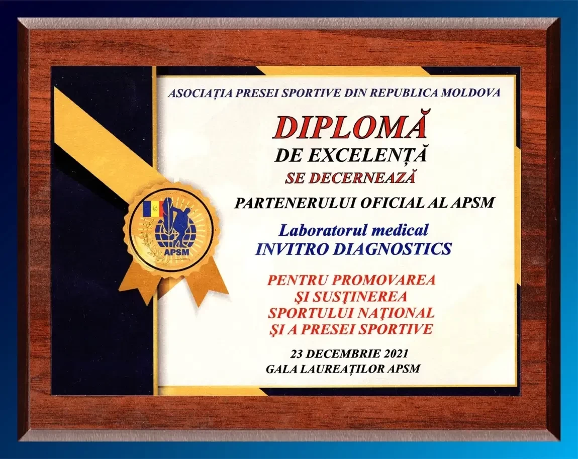
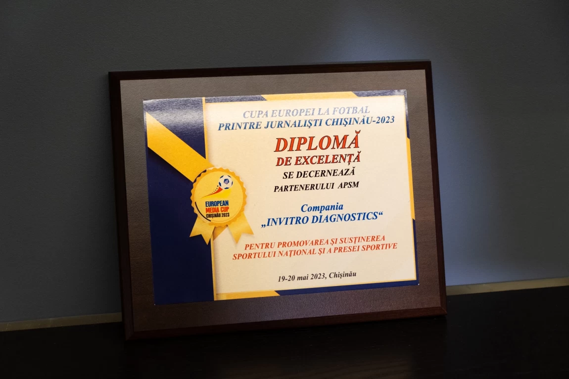
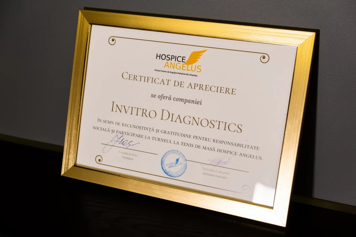
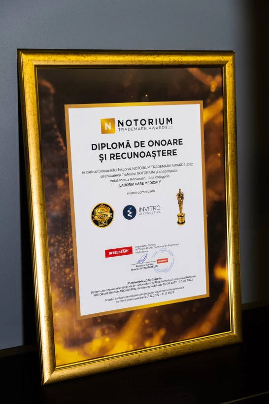
Certificates


Home collection request